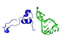
Tat (HIV)
| Tat | |||||||||
|---|---|---|---|---|---|---|---|---|---|
 | |||||||||
| 識別子 | |||||||||
| 略号 | Tat | ||||||||
| Pfam | PF00539 | ||||||||
| InterPro | IPR001831 | ||||||||
| PROSITE | PDOC00836 | ||||||||
| SCOP | 1tvs | ||||||||
| SUPERFAMILY | 1tvs | ||||||||
| |||||||||
Tat(trans-activator of transcription)は、HIV-1のtat遺伝子にコードされるタンパク質であり、ウイルスの転写効率を劇的に高める[1][2]。Tatは86–101アミノ酸からなるタンパク質であり、長さはサブタイプによって異なる[3]。TatはHIV二本鎖DNAの転写レベルを大幅に高める役割を果たす。まず二本鎖DNAから少数のRNA転写産物が産生され、そこからTatタンパク質が産生される。その後Tatは宿主因子に結合し、宿主因子によるリン酸化活性を媒介して全てのHIV遺伝子の転写を高めることで[4]、ポジティブフィードバックループを形成する。
また、TatはHIVの疾患過程においてより直接的な役割も果たしているようであり、HIV-1感染患者の血液には感染細胞から放出されたTatタンパク質が検出される[5]。TatはHIVに感染していない細胞に吸収され、非感染バイスタンダーT細胞に対し、アポトーシスを介して細胞死を誘導する毒素として直接的に作用することで、AIDSへの進行を補助する[6]。
TatはCXCR4受容体のアンタゴニストとして作用することで、感染初期にCCR5受容体を利用するビルレンスの低いM-tropic(macrophage-tropic)株の複製を選択的に促進し、M-tropic株からの変異後にのみCXCR4受容体を利用するビルレンスの高いT-tropic(T cell-tropic)株が出現するようにしているようである[5]。
機能と機構
[編集]他のレンチウイルスと同様にHIV-1はTatをコードしており、このタンパク質はウイルスゲノムの効率的な転写に必須である[7][8]。Tatは、HIV-1の新生転写産物の5'末端に位置するTAR(trans-activation response element)と呼ばれるRNAステムループ構造に結合することで作用する。TatはTARに結合することで転写複合体の性質を変化させ、CDK9とサイクリンT1からなるP-TEFb複合体(転写伸長を正に制御する)をリクルートして全長ウイルスRNAの産生を増加させる[8]。
またTatタンパク質は、プロモータークリアランス後転写伸長初期段階から全長TAR RNA転写産物が合成されるまでの間、RNAポリメラーゼII複合体とも結合している。このTatとRNAポリメラーゼIIとの相互作用は、P-TEFbには依存していない。このように転写伸長複合体にはTat結合部位が2か所あり、1つはTAR RNA、もう1つはRNAポリメラーゼII上の新生mRNA転写産物の出口付近である。このことは、1回のHIV-1 mRNA合成の促進に2分子のTatが関与していることを示唆している[9]。
In vitroでTAR特異的な結合を行うために必要最小限なTatの配列は、10アミノ酸からなる塩基性領域にマッピングされている[8][10]。この領域は大部分がアルギニンとリジン残基から構成され[8][10]、αヘリックスを形成することが示唆されている。そのため、Tatはアルギニンリッチモチーフ(ARM)RNA結合タンパク質ファミリーに属する[11]。一方、調節活性にはN末端の47残基が必要であり、この領域は転写複合体の構成要素と相互作用して転写活性化ドメインとして機能する[12]。
また、Tatは一般的ではない細胞間輸送経路を利用する。まず、Tatは細胞膜内側表面に位置するホスファチジルイノシトール-4,5-ビスリン酸(PI(4,5)P2)に高い親和性で結合する。その後、Tatは細胞膜を通過して細胞外空間に到達する。感染細胞によるTatの分泌は非常に活発であり、細胞外への搬出は産生されたTatがたどる主要な経路である[2]。
Protein transduction domain
[編集]Tatにはprotein transduction domain(PTD)と呼ばれる領域が含まれており、この領域は細胞膜透過ペプチドとしても知られている[13]。このドメインによって、Tatが細胞膜を越えて細胞内へ移行することが可能となる[14][15][16]。PTDのアミノ酸配列はYGRKKRRQRRRである[13]。このカチオン性の11アミノ酸ペプチドは、この配列を提示しているナノ粒子などの巨大分子を細胞内移行の標的とするために十分な配列である[17]。PTDポリペプチド単独でのヘパリナーゼ非依存的な移行と異なり、Tat全長のインターナリゼーションは細胞表面で負に帯電したヘパラン硫酸プロテオグリカンによって媒介される[18]。この移行過程の一部はポトサイトーシスによって媒介されている[17][19]。
PTD内には核局在シグナルGRKKRが見つかっており、さらに細胞核への移行を媒介する[19][20][21]。このドメインの生物学的役割と移行の正確な機構は未解明である[13]。
臨床的意義
[編集]Tatの阻害によるHIV感染の治療の研究が行われている[22]。HIV感染に対する治療として、Tatのアンタゴニストの利用の可能性が示唆されている[23]。
Tatタンパク質を標的としたTat Oyiと呼ばれるワクチンがBiosantechによって開発されている。このHIVワクチンは、フランスで行われた48人のHIV陽性患者を対象とした二重盲検試験で毒性を示さないことが確認された。2016年に発表された第I/IIa相研究では、調査された3種類の投与量のうちの1つでウイルスRNAの減少が示された。用量反応関係は観察されなかったため、この知見の頑健性には疑問も生じている[24]。
出典
[編集]- ^ Genes, tat - MeSH・アメリカ国立医学図書館・生命科学用語シソーラス
- ^ a b “The ins and outs of HIV-1 Tat.”. Traffic 13 (3): 355–63. (2012). doi:10.1111/j.1600-0854.2011.01286.x. PMID 21951552.
- ^ “HIV-1 TAT: Structure and Function”. Human Retroviruses and AIDS: A Compilation and Analysis of Nucleic Acid and Amino Acid Sequences. Los Alamos National Laboratory. (June 1997). pp. III-3–III-18
- ^ “Positive transcription elongation factor B phosphorylates hSPT5 and RNA polymerase II carboxyl-terminal domain independently of cyclin-dependent kinase-activating kinase”. J. Biol. Chem. 276 (15): 12317–23. (April 2001). doi:10.1074/jbc.M010908200. PMID 11145967.
- ^ a b “Selective CXCR4 antagonism by Tat: implications for in vivo expansion of coreceptor use by HIV-1”. Proc. Natl. Acad. Sci. U.S.A. 97 (21): 11466–71. (October 2000). Bibcode: 2000PNAS...9711466X. doi:10.1073/pnas.97.21.11466. PMC 17223. PMID 11027346.
- ^ “The glutamine-rich region of the HIV-1 Tat protein is involved in T-cell apoptosis”. J. Biol. Chem. 279 (46): 48197–204. (November 2004). doi:10.1074/jbc.M406195200. PMID 15331610.
- ^ “The biochemistry of AIDS”. Annu. Rev. Biochem. 60: 577–630. (1991). doi:10.1146/annurev.bi.60.070191.003045. PMID 1883204.
- ^ a b c d “NMR structure of a biologically active peptide containing the RNA-binding domain of human immunodeficiency virus type 1 Tat”. Proc. Natl. Acad. Sci. U.S.A. 91 (17): 8248–52. (August 1994). Bibcode: 1994PNAS...91.8248M. doi:10.1073/pnas.91.17.8248. PMC 44583. PMID 8058789.
- ^ “A bimolecular mechanism of HIV-1 Tat protein interaction with RNA polymerase II transcription elongation complexes”. J. Mol. Biol. 320 (5): 925–42. (July 2002). doi:10.1016/S0022-2836(02)00556-9. PMID 12126615.
- ^ a b “Trans-activation by HIV-1 Tat via a heterologous RNA binding protein”. Cell 62 (4): 769–76. (August 1990). doi:10.1016/0092-8674(90)90121-T. PMID 2117500.
- ^ “Crystal structure of HIV-1 Tat complexed with human P-TEFb”. Nature 465 (7299): 747–51. (June 2010). Bibcode: 2010Natur.465..747T. doi:10.1038/nature09131. PMC 2885016. PMID 20535204.
- ^ “Direct interaction of human TFIID with the HIV-1 transactivator Tat”. Nature 367 (6460): 295–9. (January 1994). Bibcode: 1994Natur.367..295K. doi:10.1038/367295a0. PMID 8121496.
- ^ a b c “Protein transduction: unrestricted delivery into all cells?”. Trends Cell Biol. 10 (7): 290–5. (July 2000). doi:10.1016/S0962-8924(00)01771-2. PMID 10856932.
- ^ “Delivery of bioactive molecules into the cell: the Trojan horse approach”. Molecular and Cellular Neuroscience 27 (2): 85–131. (October 2004). doi:10.1016/j.mcn.2004.03.005. PMID 15485768.
- ^ “Cellular uptake of the tat protein from human immunodeficiency virus”. Cell 55 (6): 1189–93. (December 1988). doi:10.1016/0092-8674(88)90263-2. PMID 2849510.
- ^ “Autonomous functional domains of chemically synthesized human immunodeficiency virus tat trans-activator protein”. Cell 55 (6): 1179–88. (December 1988). doi:10.1016/0092-8674(88)90262-0. PMID 2849509.
- ^ a b MacKay, J. Andrew; Szoka, Jr., Francis C. (2003). “HIV TAT Protein Transduction Domain Mediated Cell Binding and Intracellular Delivery of Nanoparticles”. Journal of Dispersion Science and Technology 24 (3): 465–473. doi:10.1081/dis-120021802. PMC 2929673. PMID 20808712.
- ^ Silhol, Michelle; Tyagi, Mudit; Giacca, Mauro; Lebleu, Bernard; Vivès, Eric (2002). “Different mechanisms for cellular internalization of the HIV-1 Tat-derived cell penetrating peptide and recombinant proteins fused to Tat”. European Journal of Biochemistry 269 (2): 494–501. doi:10.1046/j.0014-2956.2001.02671.x. PMID 11856307.
- ^ a b Eguchi, Akiko; Akuta, Teruo; Okuyama, Hajime et al. (2001). “Protein Transduction Domain of HIV-1 Tat Protein Promotes Efficient Delivery of DNA into Mammalian Cells”. Journal of Biological Chemistry 276 (28): 26204–26210. doi:10.1074/jbc.M010625200. PMID 11346640.
- ^ “Structural and functional characterization of human immunodeficiency virus Tat protein”. Journal of Virology 63 (1): 1–8. (January 1989). doi:10.1128/JVI.63.1.1-8.1989. PMC 247650. PMID 2535718.
- ^ “Mutational analysis of the conserved basic domain of human immunodeficiency virus tat protein”. Journal of Virology 63 (3): 1181–7. (March 1989). doi:10.1128/JVI.63.3.1181-1187.1989. PMC 247813. PMID 2536828.
- ^ “Recruitment of phosphatidylinositol 3-kinase to CD28 inhibits HIV transcription by a Tat-dependent mechanism”. J. Immunol. 169 (1): 254–60. (July 2002). doi:10.4049/jimmunol.169.1.254. PMID 12077252.
- ^ “4-Phenylcoumarins as HIV transcription inhibitors”. Bioorg. Med. Chem. Lett. 15 (20): 4447–50. (October 2005). doi:10.1016/j.bmcl.2005.07.041. PMID 16137881.
- ^ “Intradermal injection of a Tat Oyi-based therapeutic HIV vaccine reduces of 1.5 log copies/mL the HIV RNA rebound median and no HIV DNA rebound following cART interruption in a phase I/II randomized controlled clinical trial”. Retrovirology 13: 21. (April 2016). doi:10.1186/s12977-016-0251-3. PMC 4818470. PMID 27036656.
